Major Diseases of Mushroom
-
Fungal Diseases
-
Bacterial diseases
-
Viral diseases
1.Fungal Diseases
Dry Bubble
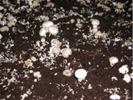
- Verticillium fungicola
- Muddy brown, often sunken spots on the cap of the mushrooms
- Greyishwhite moldy growth seen on pileus
- Later stage mushroom becomes dry and leathery
- Initially infected one are not develop or remain small
- Main source of infection
- Debris
- Dust on floors of growing house
- Spread
- Water splashes on healthy mushroom
- Sciarid and phorid flies over long distance
- Favorable temperature 28o c
- Poor ventilation
- High humidity
Management of Dry Bubble
- Pick and destroy infected mushroom to prevent spread
- Sanitary conditions in growth house
- Lower the temperature to 14oc when disease noticed
- Use clean equipment
- Control flies and mites
- Bubble can destroy with salt
Management by using salt
- Use of clean pasteurized casing media
- Properly maintained air filtering system.
- Water early infection centers with formaldehyde
- For protective measure use zineb
- Apply chlorothalonil at casing or mix into casing material
Dry bubble in Oyster

False Truffle
- Diehliomyces microsporus
- Competitor than a pathogen
- Appears as cottony weft of mycelium on bed surface
- Wefts turn to dense small reddish brown,wrinkled,stromatic bodies resemble a truffle
- Infected bed have peculiar disagreeable odour
- Reduced yield at mycelia exist
- Introduced through soil
Management
- Good sanitation
- Proper Pasteurization of casing material
- Low temperature during spawn run
Wet Bubble
Mycogone perniciosa
- Malformed mushrooms with swollen stipes
- Reduced or deformed caps
- Undifferentiated tissue becomes necrotic and a wet, soft rot emit bad odor
- An amber liquid appears on infected mushrooms.
- Mushrooms become brown in color
- Bubbles may be as large as a grapefruit.
- The fungus is spread via airborne dust and contaminated casing.
- It is also a parasite of wild mushrooms.
- It produces two spore types,
- one which is small and water-dispersed like Verticillium,
- second which is a large resting spore capable of persisting for a long time in the environment
Control
- Sanitation in growth house
- Clean environment around cultivation area
- Incorporating benzimidazoles in the casing.
- Benomyl at the rate of 0.95 g/m2,
- Carbendazim and thiabendazole at the rate of 0.62 g/m2
Cobweb
- Cladobotryum dendroides
- White silky growth grows over surface of casing soil
- It climb up and cover mushrooms comes in it’s path
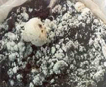
Older mycelium changes from silky to granular white
- Infected mushroom become soft
- Later engulfed by cottony ball of mycelia
- Serious problem where year around growing is practiced
- Cobweb mold is darker than mycelium... almost grey as compared to white.
- Main source of infection is casing soil
- A cottony mycelium grows over casing
- the mycelium soon envelopes the mushroom with a soft mildewy mycelium and causes a soft rot.
- It is also a parasite of wild mushrooms.
- Cobweb mold is favored by high humidity.
Management of Cobweb
- Identify disease symptoms early, not only the web but also cap spotting.
- Treat spotty infections with a alcohol drenched paper towel
- Cover infected areas with salt
- Change from light peats to heavy peat casing may encourage disease development, but heavy black peats are not responsible for initial infections.
- Heavier casing may require increased water applications, therefore may encourage the spread and development the disease.
- Heavily infected 2nd or early 3rd breaks should be steamed off to reduce the spore load on the farm.
- Control strategies include lowering humidity and /or increasing air circulation
- Increase hygiene of the harvesting and watering department.
- Judicious applications of Benzimidazole fungicides should be made
- Chlorothalonil should be included in the fungicide application program
Common chemicals used to control diseases in mushroom
Fungicide |
Against Pathogen |
Doze |
| Benomyl and Carbendazim |
Dactylium, Mycogone, Trichoderma, Verticillium |
Mix 240 g/100m2
With casing or in water @ 240g/200 l/100m2 |
| Chlorothalonil |
Mycogone, Verticillium |
200 ml/200 l/100m2
1st spary 1 week after casing & 2nd after 2 week |
| Prochloroz manganese |
Dactylium, Mycogone, Verticillium |
300g/ 100 l/ 100 m2 for single spary
Or 113 g/100 l/100 m2 if 2 spray |
| Thiabendazole |
Dactylium, Mycogone, Verticillium |
Mix 240 g/100m2
With casing or in water @ 240g/200 l/100m2 |
| Zineb |
Dactylium, Mycogone, Verticillium |
350 g/ 100m2, (dust) every week after casing.
1 kg/1000 l @ of 5 l/10m2 after casing & between flushes |
Bacterial Spot/Pit /Brown Blotch
- Pseudomonas tolaasii.
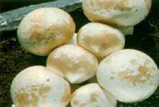
- Pale yellow spots on the surface of the piles later it turns to yellow
- In Sevier case mushrooms are radially streaked
- Damage at storage and transit

- Source of contamination may be soil or water
- High humidity and watery conditions are favorable for disease
- Vector: Tryoglyphid mite
- Lesions on tissue that are pale yellow initially, later become a golden yellow or rich chocolate brown.
- Discoloration is superficial (not more than 2 to 3 mm)
- Underlying tissue may appear to be water soaked and grey.
- Blotches appear in early button stage,
- Appear on any age - even on harvested refrigerated mushrooms
- At favorable moisture conditions spots enlarge and coalesce, sometimes covering entire cap
- Mushroom stems can also be blemished similarly
- Typical spotting is observed at or near the edge of mushroom caps wherever caps remain wet for a period of 4 to 6 hours or longer after water has been applied
- If very dry conditions occur after blotch has developed, infected caps may crack radially as the mushroom expands
- Casing and air-borne dust are primary source.
- The bacterial pathogen is probably present in most casing material, even after pasteurization.
- Occurrence of disease associated with the size of the bacterial population on the mushroom cap, rather than on the population in the casing, which explains why a prolonged wet period on the cap precedes disease occurrence.
- Spread by splash, tools, flies and nematodes.
- Moisture content of less than 62 percent at spawning preconditions mushrooms to blotch infection.
Management
- Sanitation
- Lowering humidity
- Watering with a 150 ppm chlorine solution (calcium hypochlorite products are used since sodium hypochlorite products may burn caps).
- If the mushroom stays wet, however, chlorine has little effect since the bacterial population reproduces at a rate that neutralizes the effect of the oxidizing agent.
Mummy disease
- The disease was first described in 1942 by CM Tucker and JB Routien in the United States

- Fruit bodies have tilted caps,
- Early veil breaking,
- Base of the stem enlarged
- Curved stalk
- Tissue of the mushroom becomes
- Spongy,
- Dry and
- Brown
- Mummified appearance.
- Rapid rate of spread through the bed, up to 30cm (12 inches) daily.
- Infected mushroom is tough and dry texture
- Gritty texture appeared when cut.
- Water-soaked appearance and cavities in the mushroom tissue
Management
- Dig a trench to separate the diseased area from the healthy.
- Dug around 2m (6-8ft) ahead of the advancing disease and the trench itself to be several inches wide.
- All compost and casing has to be removed from the trench
- Gap thoroughly disinfected.
- If crop is growing in separate containers, the usual advice is to isolate or dispose of them.
- Good hygiene is strongly recommended.
- Virus (several)
- Double-stranded RNA
- Reduced cropping,
- bare patches on the beds,
- long-bent stalks with small caps,
- Premature opening of mushrooms,
- Stalks tapering towards the base of stalk,
- Dying pinheads
- Infected mycelium grows slowly in the beds and fruiting bodies are not produced.
- Infection of the crop at spawning lead to a higher level of disease
Spread and source of infection of virus
- Infected mushroom spores
- Mycelium from previous crops also survive in the trays
- Mushroom sheds can also release infected spores
- Dust from around the farm may introduce infected spores
- Only 10 infected spores are required for a disease outbreak.
- Farm hygiene
- Maintain 60oC temperature throughout the compost
- Filter air and seal rooms properly to prevent spores from entering during cool down phase of compost.
- Clean equipments
- Ensure workers have clean-spare clothes
- Ensure absolute filters are fitted to spawn-run buildings
- Clean trays to prevent infection from old-infected mycelia
Green moulds
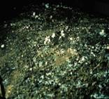
- Trichoderma koningii
- T.viride
- T.aggressivum f.sp.aggressivum
- Dark green mould patches on casing spreading to lesions on stems.
Control of green mould
- Sanitation and hygiene programme, especially targeting post crop
- Cover spots with sodium hypochlorite solution, salt, lime or gypsum and lime mix.
- Good insect and mite control
- Personnel movement patterns further reduce the spread of the disease.
- chlorothalonil at casing or mix into casing material 254 mL formulation per 100 m2 of production
- Chlorothalonil is not effective against an established infection but lowering the infection
Cinnamon Mould
- Chromelosporium fulva (Peziza ostrachoderma)
- The color of this mold ranges from yellow gold to golden brown to cinnamon brown.
- It grows rapidly in circular patches.
- It is very common in soil, and flourishes on damp wood.
- Areas in compost overheated during spawn run may be colonized.
- Improperly conditioned compost will also support growth,
- It often occurs on sterilized soil.
- Sexual fruiting bodies may appear several weeks after the first appearance of the mold.
- Spores are airborne
Pink Mold
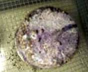
- Neurospora spp.
- Commonly to occasionally seen on agar and grain.
- It is ubiquitous in nature, occurring on dung, in soils and on decaying plant matter.
- Neurospora spores germinate more readily at elevated temperatures.
- The pink mold seen in mushroom culture is most frequently Neurospora sitophila, a pernicious contaminant that is difficult to eliminate.
- All infected cultures should be removed as soon as possible from the laboratory and destroyed.
- A thorough cleaning of the laboratory is absolutely necessary.
- If contamination persists, remove all spawn and start anew
Inky Cap
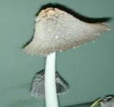
- Coprinus spp.
- These are evidence of free ammonia in the compost.
- Their delicate gray caps autodigest quickly.
- Inky caps are indicators of nitrogen over supplementation or a poorly managed Phase II compost.
- If there is too much residual ammonia, Phase II thermophilic microflora may be unable to convert all the ammonia into microbial protein.
- fungus is strongly cellulolytic.
|


